Home
/ Leg Anatomy Muscles Ligaments And Tendons - Where Or Where Has My Patella Gone Direct Orthopedic Care : The leg anatomy includes the quads, hams, glutes, hip flexors, adductors & abductors.
Leg Anatomy Muscles Ligaments And Tendons - Where Or Where Has My Patella Gone Direct Orthopedic Care : The leg anatomy includes the quads, hams, glutes, hip flexors, adductors & abductors.
Leg Anatomy Muscles Ligaments And Tendons - Where Or Where Has My Patella Gone Direct Orthopedic Care : The leg anatomy includes the quads, hams, glutes, hip flexors, adductors & abductors.. Muscles, tendons, and ligaments run along the surfaces of the feet, allowing the complex movements needed for motion and balance. In human anatomy, there are several strong bands of connective tissues called ligaments, which hold the bones of the ankles together. The muscles, tendons, and ligaments that support the ankle joint work together to propel the body. The journal no longer participates in pmc. These all work together to bear weight.
Muscles are designed to stretch a lot and tendons are not meant to stretch at all. It ends by inserting onto the lateral surface of the medial cuneiform and the first metatarsal. Medical project (bone, muscles, ligaments and joints bio mechanics) am working on. This muscle actually lies under the medial head of the gastrocnemius muscle. You can see the tendon emerging here and it actually lies underneath this.
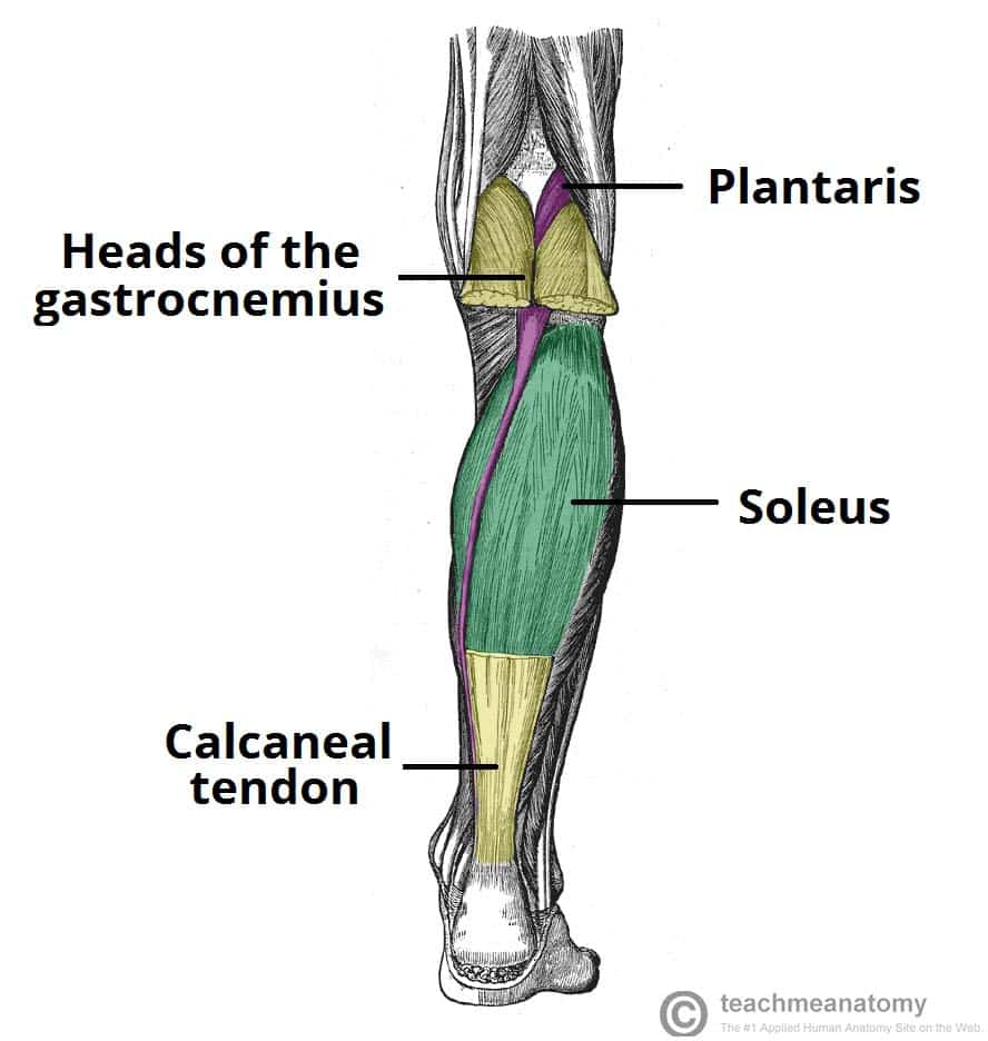
Muscles Of The Leg Anterior Lateral Posterior Teachmeanatomy from teachmeanatomy.info Tendons consist of densely packed collagen fibers. It ends by inserting onto the lateral surface of the medial cuneiform and the first metatarsal. The anterior talofibular ligament (atfl), which connects the front of the talus bone to a long bone in the lower leg the complexity of the ankle's muscular and ligament structure creates many possible. Muscles, either individually or in groups, are supported by fascia. Learn about the muscles, tendons, bones, and ligaments that comprise the knee joint anatomy. Tendons are the connective tissue that connects our muscles to the bones and just like the ligaments they are made of collagen. There are four muscles in the anterior compartment of the leg. Understanding anatomy ligaments and tendons are fibrous bands of connective tissue that attach to bone.
Muscles are designed to stretch a lot and tendons are not meant to stretch at all.
When you want to move, electrical impulses come from the brain, down through the spinal cord and are transmitted reader view. Ligaments also support the lower end of the leg where it forms a hinge for the ankle. In other words, this page excludes information about the calf muscles… The achilles tendon connects the heel to the calf muscle and is essential for running, jumping, and standing on the toes. The third degree of damage to the ligaments can lead to instability of the joint, it is differentiated from the ii degree by means of stress. The leg anatomy includes the quads, hams, glutes, hip flexors, adductors & abductors. Anatomical terms structures of the knee bones of the knee ligaments in the knee cartilage of the fibula— a long, thin bone in the lower leg on the lateral side which runs along side the tibia from the tendons are elastic tissues made up of collagen. Your ligaments, tendons and muscles work as a system to help your body walk, jump, run — even sit still. Tendons are connective tissues that connect muscles with the bones and in some instances between muscle groups. One way our muscles work: Learn the origin/insertion, functions & exercises for the specifically, this page discusses all the major muscle groups of the upper leg. The system of ligaments in the vertebral column, combined with the tendons and muscles, provides a natural brace to help protect the spine from injury. Unlike tendons, which connect muscle to bone, ligaments connect bones to other bones.
The human leg, in the general word sense, is the entire lower limb of the human body, including the foot, thigh and even the hip or gluteal region. Ligaments, muscles and tendons keep us connected and help us move. One way our muscles work: Muscles, ligaments, & tendons by: The achilles tendon connects the heel to the calf muscle and is essential for running, jumping, and standing on the toes.
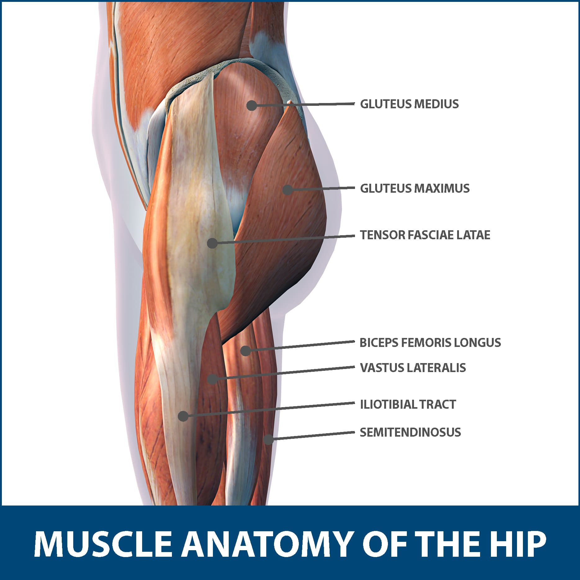
Hip Muscle Strains Info Florida Orthopaedic Institute from www.floridaortho.com The quadriceps muscle and tendon extend the lower leg and play an important role in patellar distally, the biceps muscle joins the lateral collateral ligament and forms a conjoined tendon that popliteus muscle and arcuate ligament in a 40 year old male. Unlike tendons, which connect muscle to bone, ligaments connect bones to other bones. Tendons consist of densely packed collagen fibers. Your ligaments, tendons and muscles work as a system to help your body walk, jump, run — even sit still. Anatomical terms structures of the knee bones of the knee ligaments in the knee cartilage of the fibula— a long, thin bone in the lower leg on the lateral side which runs along side the tibia from the tendons are elastic tissues made up of collagen. There are four muscles in the anterior compartment of the leg. Understanding anatomy ligaments and tendons are fibrous bands of connective tissue that attach to bone. Medical project (bone, muscles, ligaments and joints bio mechanics) am working on.
Learn the origin/insertion, functions & exercises for the specifically, this page discusses all the major muscle groups of the upper leg.
Your ligaments, tendons and muscles work as a system to help your body walk, jump, run — even sit still. Collectively, they act to dorsiflex and invert the foot at the ankle joint. Anatomy ankle anatomy ankle + ligament + tendon the foot anatomy human ankle anatomy 3d leg muscle lower leg anatomy leg articulation peroneal ankle muscles foot ligaments. Katelyn forsee how do our muscles work? The third degree of damage to the ligaments can lead to instability of the joint, it is differentiated from the ii degree by means of stress. In human anatomy, there are several strong bands of connective tissues called ligaments, which hold the bones of the ankles together. Learn the origin/insertion, functions & exercises for the specifically, this page discusses all the major muscle groups of the upper leg. Ligaments also support the lower end of the leg where it forms a hinge for the ankle. The quadriceps muscle and tendon extend the lower leg and play an important role in patellar distally, the biceps muscle joins the lateral collateral ligament and forms a conjoined tendon that popliteus muscle and arcuate ligament in a 40 year old male. Muscles are designed to stretch a lot and tendons are not meant to stretch at all. The tendons of the edl can be palpated on the dorsal surface of the foot. Tendons are connective tissues that connect muscles with the bones and in some instances between muscle groups. The patellar tendon on the front of the knee is part of the quadriceps mechanism.
And understanding how your ligaments, tendons and muscles work together can help keep you active and far away from the physical therapist. Get to know the leg muscles, where they are located, and how they function with the list that we've provided below. Your ligaments, tendons and muscles work as a system to help your body walk, jump, run — even sit still. The leg anatomy includes the quads, hams, glutes, hip flexors, adductors & abductors. Tendons are the connective tissue that connects our muscles to the bones and just like the ligaments they are made of collagen.
Plantaris Muscle Wikipedia from upload.wikimedia.org The achilles tendon connects the heel to the calf muscle and is essential for running, jumping, and standing on the toes. Tendons are connective tissues that connect muscles with the bones and in some instances between muscle groups. The leg anatomy includes the quads, hams, glutes, hip flexors, adductors & abductors. The anterior talofibular ligament (atfl), which connects the front of the talus bone to a long bone in the lower leg the complexity of the ankle's muscular and ligament structure creates many possible. Unfortunately many of us live in a bodily environment where ligaments tend to be overstretched or lax in tone. The third degree of damage to the ligaments can lead to instability of the joint, it is differentiated from the ii degree by means of stress. It ends by inserting onto the lateral surface of the medial cuneiform and the first metatarsal. Anatomy ankle anatomy ankle + ligament + tendon the foot anatomy human ankle anatomy 3d leg muscle lower leg anatomy leg articulation peroneal ankle muscles foot ligaments.
In human anatomy, there are several strong bands of connective tissues called ligaments, which hold the bones of the ankles together.
Muscles, ligaments, & tendons by: Tendons consist of densely packed collagen fibers. The muscles, tendons, and ligaments that support the ankle joint work together to propel the body. They are the continuations of muscles and. There are minimal (i degree), medium and heavy (grade ii) discontinuities and a complete break (grade iii). The patellar tendon on the front of the knee is part of the quadriceps mechanism. The journal no longer participates in pmc. Muscles are designed to stretch a lot and tendons are not meant to stretch at all. Unfortunately many of us live in a bodily environment where ligaments tend to be overstretched or lax in tone. The system of ligaments in the vertebral column, combined with the tendons and muscles, provides a natural brace to help protect the spine from injury. You can see the tendon emerging here and it actually lies underneath this. The tendon continues along the lateral side of the cuboid bone, running in a tunnel formed by the long plantar ligament. The tissues that are included in sclerotogenous pain include: